Friday, December 21, 2018

Regorafenib for patients with hepatocellular carcinoma who progressed on sorafenib treatment (RESOURCE): a randomized, double-blind, placebo-controlled, phase 3 trial


Summary:


Hepatocellular carcinoma (HCC) locoregional treatment includes surgical resection, transplantation, ablation and chemoembolization. For those patients who are not candidates for such treatment, medical management with the oral multikinase inhibitor sorafenib, can provide improvement in overall survival. This study aimed to evaluate the safety and efficacy of regorafenib, for patients in whom HCC has progressed during treatment with sorafenib.

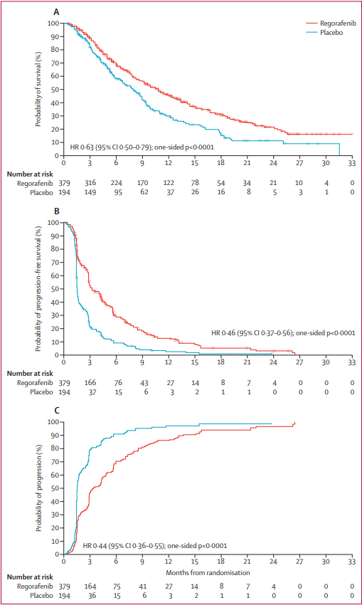
Across 152 centers and 21 countries, 573 patients with progression of HCC on sorafenib were enrolled in a randomized 2:1 regorafenib to placebo, double-blind, placebo-controlled phase 3 clinical trial. The primary endpoint was overall survival analyzed by intention to treat. Median overall survival was 10.6 months (95% CI 9.1 – 12.1) with regorafenib and 7.8 months (6.3 – 8.8) with placebo. Median progression-free survival was 3.1 months (2.8 – 4.2) with regorafenib and 1.5 months (1.4 – 1.6) with placebo by mRECIST criteria; median time to progression of 3.2 months (2.9 – 4.2) and 1.5 months (1.4 – 1.6) respectively. The safety profile of regorafenib in HCC is consistent with the use of this medication in other malignancies. The most common reason for discontinuation of regorafenib was disease progression (60%, 83% in the control group).

This study concludes that regorafenib provides a clinical improvement in overall survival in patients with progression of HCC while on sorafenib. Two patients had a complete tumor response by mRECIST criteria. This suggests that the sequential use of two multikinase inhibitors with partially overlapping target profiles can provide benefit to this patient population.


Figure 2. Kaplan-Meier analysis of overall survival (A), progression-free survival (mRECIST; B), and time to progression (mRECIST; C)

Commentary:


Regorafenib offers a viable option for patients who have failed sorafenib and has the potential to add to the current treatment armamentarium. Multiple clinical trials with second line therapies including TACE (Kudo et al., GIDEON trial, Zhang et al.), HAIC (SIRIUS trial, Terashima et al., Shao et al.) and alternative medications (Llovet et al., Zhu et al.) have demonstrated variable benefit depending on the treatment timing or drug.

The STAB Study Phase II data, recently published by Sato et al. indicates a similar class of patients (Child-Pugh class A with Barcelona Stage C disease) may benefit from concurrent TACE with sorafenib therapy with a mean overall and progression free survival of 17.3 and 5.4 months respectively, longer than those with regorafenib therapy. While this patient population was chemotherapy-naïve, it offers a potential treatment option for those patients on sorafenib prior to treatment failure. The introduction of regorafenib has the potential to change IR treatment for refractory HCC as concurrent regorafenib-TACE or alternative IR therapies may become a viable option in the future.


Bruix J, Qin S, Merle P, et al. Regorafenib for patients with hepatocellular carcinoma who progressed on sorafenib treatment (RESOURCE): a randomized, double-blind, placebo-controlled, phase 3 trial. Lancet. 2017; 389: 56-66.

Post Author:
Nicole A. Keefe, MD
Resident Physician
Department of Radiology and Medical Imaging
University of Virginia
@NikkiKeefe

Friday, December 14, 2018

Patient and Facility Demographics Related Outcomes in Early-Stage Non-Small Cell Lung Cancer Treated with Radiofrequency Ablation: A National Cancer Database Analysis 


Summary


The role of patient demographics and facility volume play with regards to survival after surgery for NSCLC has been established in multiple prior studies. However, while survival data is available for radiofrequency (RF) ablation of NSCLC, the impact of patient demographics and facility volume is not known in these patients undergoing RF ablation.

The purpose of this investigation was to evaluate the effect that these factors have on OS after RF ablation in early-stage NSCLC using the National Cancer Database (NCDB). The investigators performed a retrospective cohort study of all patients diagnosed with NSCLC from 2004 to 2014. Patients with metastatic and or nodal disease were excluded (T1aN0M0 and T1bN0M0). Additionally, patients who received chemotherapy or radiation at any time were excluded. The final cohort was a total of 967 patients. Patient demographic factors incorporated into the analysis included insurance status, income, education level and residence type. Facility demographic variables evaluated included facility type (community cancer program, comprehensive community cancer program, academic/research program, or integrated network cancer program) and facility volume. Patients were separated into those treated at high-volume centers (HVCs) and those who were not (non-HVC). After propensity score-weighted analysis of a matched cohort of 574 patients, the significant improvement in OS persisted with 1-, 3-, and 5-year OS of 89.4%, 51.2%, and 27.7%, respectively, in the HVC group, compared to 85.2%, 42.0, and 19.6%, respectively, in the non-HVC group (P=.015). Worse OS after RF ablation was significantly associated with increasing age and higher T-classification.


Commentary


This study suggests there is a significant survival benefit in patients treated with RF ablation for NSCLC in higher volume institutions. What to do with this knowledge remains the question. Additionally, more work will need to be done to look further at the specific variables accounting for this difference, apart from the higher volumes done at these centers. With all things equal, ablative technologies for NSCLC can then be pitted head to head with the current standard of care.

Click here for abstract

Lam, Alexander, et al. Patient and Facility Demographics Related Outcomes in Early-Stage Non-Small Cell Lung Cancer Treated with Radiofrequency Ablation: A National Cancer Database Analysis. Journal of Vascular and Interventional Radiology. November, 2018. Volume 29, Issue 11, 1535–1541.

Post Author: 
Zagum Bhatti, MD
Assistant Professor
Department of Radiology, Interventional Radiology Division
University of Texas Health Science Center at Houston, Houston, TX
@ZagumBhatti

Thursday, December 13, 2018

From the SIR Residents and Fellows Section (SIRRFS) Teaching Topic: Uterine Artery Embolization for the Treatment of Adenomyosis: A Systematic Review and Meta-Analysis


de Bruijn AM, Smink M, Lohle PN, Huirne JA, Twisk JW, Wong C, Schoonmade L, Hehenkamp WJ. Uterine Artery Embolization for the Treatment of Adenomyosis: A Systematic Review and Meta-Analysis. Journal of Vascular and Interventional Radiology. 2017 Oct 9.

Click here for abstract

This evidence-based review article evaluated the effect of uterine artery embolization (UAE) on symptom reduction in adenomyosis with respect to length of follow-up (< 12 months or >12 months) and whether patients had pure (adenomyosis alone) or combined (adenomyosis and fibroids) disease. The meta-analysis supported that uterine artery embolization (UAE) for both pure and combined adenomyosis resulted in significant short-term and long-term symptom improvement. There were limitations in that no randomized controlled trials were available, many included studies were retrospectively evaluated, many studies had a small sample size, and many studies offered vague description of methodology. As well, the sample size of most included studies was not sufficient to draw solid conclusions. This called for future trials to use standardized outcomes and validated questionnaires to render outcomes that can be extrapolated in systematic reviews.


Figure. Improvement of clinical symptoms (%).

Clinical Pearls


What is the pathophysiology of adenomyosis?

Adenomyosis defined as heterotopic endometrium in the myometrium and has diffuse, segmental, superficial, and deep forms. It is typically considered a disease of multiparous women in the late reproductive years. Though the etiology is still not well known, adenomyosis is thought to occur from either endomyometrial invagination of the endometrium or de novo from mullerian rests. There is also evidence that the junctional zone (JZ), which separates the subendometrial myometrium from the outer myometrium (appears as a dark band on T2WI), plays a role, as disease in the JZ predisposes to secondary infiltration of endomyometrial elements. Aside from older, multiparous women, JZ distortion can also be found in young, nulliparous women with heavy periods. A JZ > 12 mm is highly predictive of disease and < 8 mm essentially excludes it. Six studies in the meta-analysis reported a JZ reduction of 13.7%-38% after UAE. Smeets et al reported a thicker JZ at baseline to be a possible predictor of UAE failure in patients with adenomyosis. Patients who underwent hysterectomy owing to persisting symptoms compared with patients with clinical improvement showed a statistically significant thicker JZ at baseline (P = .028) and during 3 months (P = .034) of follow-up. Nijenhuis et al reported the mean JZ thickness at baseline not to be significantly thicker in groups with insufficient response (P = .17); however, it was statistically significant thicker in 4 patients needing additional therapy (P = .004).

What other gynecological pathologies are associated with adenomyosis?

Concurrent benign, estrogen-dependent gynecologic pathology, namely leiomyoma and endometriosis, is often present, increasing operative risks for major gynecologic surgery. There is also a known association between recurrent fetal loss and adenomyosis, though there is no direct evidence that adenomyosis is a risk factor for spontaneous abortion. The authors of this study state that in addition to the use of standardized outcomes and validated questionnaires, future trials should focus on fertility results in patients with symptomatic adenomyosis. Only two of the thirty-four studies in their meta-analysis reported fertility outcomes in terms of pregnancy outcome after UAE. Kim et al reported on 5 pregnancies. Two of these patients underwent elective abortion owing to unwanted pregnancies. The remaining three patients carried to term and showed no signs of uteroplacental vascular insufficiency or abnormal uterine contraction during labor or postpartum. The pregnancies resulted in 2 vaginal deliveries and 1 elective cesarean delivery because of previous cesarean delivery. The average weight of neonates was 3.2 kg (range, 3.1–3.4 kg). Yao et al described 1 pregnancy after UAE reporting termination of the pregnancy after 26 weeks. The reason for termination was not reported. Of 99 patients, 6 (6%) became pregnant after UAE. There is a paucity of high-quality data documenting the ability to conceive following UAE. Studies have reported between 48-59% live birth pregnancy rates, although the data is limited and influenced by age and current fertility status. Based on limited data, a discussion should take place with the patient about her fertility goals and a referral to a reproductive endocrinologist may also be helpful.

Why are symptoms of adenomyosis?

Though adenomyosis may be asymptomatic, it typically produces the symptoms of dysmenorrhea, dyspareunia, and chronic pelvic pain. These are also the most common symptoms of the other estrogen-dependent, benign gynecologic pathologies of endometriosis, leiomyoma, and endometrial polyp, which makes delineation of the symptoms profile difficult if the processes are concurrent. The included studies in this meta-analysis reported improvement of clinical symptoms in 83.1% (872 of 1,049) of patients. Only five of the thirty-four studies used the standardized Uterine Fibroid Symptom and Quality of Life (UFS-QOL; Spies JB, Coyne K, Guaou Guaou N, Boyle D, Skyrnarz-Murphy K, Gonzalves SM. The UFS-QOL, a new disease-specific symptom and health-related quality of life questionnaire for leiomyomata. Obstet Gynecol 2002; 99:290–300) questionnaire to evaluate health-related quality of life (HRQOL) and symptom severity. This use of this questionnaire should be used in future trials to render outcomes that can be extrapolated in systematic reviews.

Question to Consider


What are complications of UAE?

There were 20 studies that reported on complications in 615 patients. Abdominal pain occurring directly following the procedure until 2 weeks after the procedure was described in 361 of 413 patients (87.4%). Persistent amenorrhea was reported in 28 of 445 patients (6.3%) in 13 studies. All of these patients were > 40 years of age. A pseudoaneurysm was reported, which was treated with a thrombin injection. Spontaneous expulsion of leiomyomata was reported in ten patients. Endometritis was suspected in four patients. These patients were treated with broad-spectrum antibiotics. Deep venous thrombosis of the calf occurred in one patient; however, this patient did not require anticoagulation therapy. No deaths or other complications occurred.

For pain control, patient controlled analgesia with IV morphine, meperidine, hydromorphone, or fentanyl is effective. Some interventionalists prefer epidural or oral administration of narcotics. UAE patients often suffer nausea post-procedure and require either prophylactic administration or, as needed, delivery of antiemetic such as odansetron. Anti-inflammatory agents such as IV ketorolac or oral ibuprofen periprocedurally or postoperatively are commonly used. Some physicians prefer administering steroids pre-procedurally, as well, to help limit post-embolization symptoms.

The risk of infection in UAE is low with reported rates of 0.2-1% of patients. Infection usually occurs in a delayed fashion, one to two weeks after the procedure. There have been several cases of fatal sepsis occurring after UAE in patients who did not receive prophylactic antibiotics. Currently, SIR recommends prophylaxis with a single dose of 1 mg Cefazolin IV as a common choice to cover typical organisms causing infection, including skin flora and E. Coli

1. Genc M, Genc B, Cengiz H. Adenomyosis and accompanying gynecological pathologies. Arch Gynecol Obstet 2015; 291:877–881.
Brosens JJ, de Souza NM, Barker FG. Uterine junctional zone: function and disease. Lancet 1995; 346:558.

3. Spies JB, Coyne K, Guaou Guaou N, Boyle D, Skyrnarz-Murphy K, Gonzalves SM. The UFS-QOL, a new disease-specific symptom and health-related quality of life questionnaire for leiomyomata. Obstet Gynecol 2002; 99:290–300.

4. LoVerme W, James M, Ho Ahn S. “Patient Evaluation: Genitourinary System.” Patient Care in Vascular and Interventional Radiology. Third Edition. Society of Interventional Radiology, 2016. 99-111. Print.

Post Author:
Rajat Chand
Radiology Resident
John H. Stroger, Jr. Hospital of Cook County
Wednesday, December 12, 2018

Outcomes of Surgical Resection after Radioembolization for Hepatocellular Carcinoma


Summary


This is a retrospective study evaluating all patients who underwent both radioembolization (TARE) and subsequent hepatic resection in the setting of hepatocellular carcinoma (HCC). The study population (31 patients) was derived from 155 patients with HCC who underwent TARE but were also potential surgical resection candidates [preserved liver function, unilobar disease (or bilobar within Milan), no portal vein thrombus, no metastases]. The remaining patients, of the 155, were either transplanted (33) or considered for transplant (91).

The study population was made of 94% CP A and 77% BCLC Stage A disease. Technique of TARE was either lobar at a median dose of 128 Gy or segmental ablative at a medium dose of 254 Gy. 27/31 patients had a single treatment. There were no radiation-induced complications.

In addition to surgical outcomes, the authors looked extensively at the future liver remnant (FLR) following Y-90 therapy, as 25/31 patients underwent a major hepatic resection. FLR was calculated from the pre-TARE to the pre-surgical imaging. FLR increased significantly from 35% to 45% in those who received lobar Y-90 (20/25) and from 32% to 34% in those who received segmentectomy (5/25).

EASL response rates after TARE were evaluated. Stable disease was present in 39%, partial response 29%, and complete response 29%. Surgical resection after TARE occurred a median of 2.9 months after initial therapy. 24/31 patients had > 90% necrosis based on pathologic specimens.

In follow-up, 9/31 patients developed either a systemic or liver recurrence (all of whom had primary tumors > 5 cm), preferentially so in those who were EASL stable disease or had less pathologic necrosis. These findings yielded hazard ratios to develop recurrence for EASL responders of 0.18 (CI 0.5-0.7) and for 50-99% pathologic necrosis patients of 0.12 (CI 0.02-0.6). Median time to recurrence was 34 months. Median survival time was 96% at 1 and 2 years in the setting of a median follow-up time of 13 months.


Commentary


From a large number of TARE patients, this study was able to parse out a subset who underwent both TARE and subsequent surgical resection. The authors successfully demonstrate acceptable outcomes of surgical resection in the setting of prior radioembolization. They also effectively elucidate that response after Y-90 therapy by both imaging and pathologic criteria is a harbinger of future risk of recurrence after surgery.
Indirectly, this article seems to also be about the use of radioembolization to increase FLR, thus allowing surgical resection. The authors mention the potential benefits of this use, i.e. allowing local tumor control and mitigating the potential increase in local tumor growth, should one use for example portal vein embolization.
Currently TARE is often utilized in the setting non-operative HCC candidates. The authors expanded its use in this study, it seems using it as a potential bridge to resection for those in whom the FLR was insufficient to allow for surgical resection. Directly addressing this potential use of Y-90 would be helpful in future studies, as the current data only reflects the outcomes of those who successfully underwent surgery. Indeed, perhaps the success of this procedure in terms of FLR growth is much lower than theorized.

Click here for abstract

Gabr A, Abouchaleh N, Ali R, et al. Outcomes of surgical resection after radioembolization for hepatocellular carcinoma. J Vasc Interv Radiol. 2018; 29(11): 1502-1510.

Post Author:
Daniel P. Sheeran, MD
Assistant Professor
Department of Radiology and Medical Imaging
Division of Vascular and Interventional Radiology
University of Virginia

Tuesday, December 11, 2018

Diagnostic accuracy of noncontrast MR angiography protocols at 3T for the detection and characterization of lower extremity peripheral arterial disease


Summary


A clinical study evaluating non-contrast magnetic resonance angiography (MRA) to gadolinium (Gd)-based contrast MRA for lower extremity peripheral artery disease (PAD) in 20 patients was performed at 3T (vs. 1.5 T as most published diagnostic accuracy studies). Two established non-contrast MRA protocols were compared to conventional Gadolinium (Gd)-contrast enhanced MRA: (1) Quadruple inversion-recovery (QIR) Electrocardiogram combined with (ECG)-gated fast spin echo (ECG-FSE) and (2) quiescent-interval single-shot (QISS).

Imaged patients (n = 20) had intermittent claudication (Fontaine stage II) with an ankle-brachial index of < 0.9. Exclusion criteria included rest pain, nonhealing ulcers, estimated glomerular filtration rate < 30 mL/min/1.73 m2, pregnancy, claustrophobia, clinical instability or any contraindication to 3T MR imaging.

In this study, imaging datasets were reviewed in a blinded fashion by two radiologists. Non-Gd protocols demonstrated comparable diagnostic accuracy with high specificity compared to Gd-MRA protocols.


Commentary


The authors present a well-designed study evaluating forthcoming techniques in non-contrast enhanced MRA for the evaluation of lower extremity peripheral vascular disease. The techniques demonstrate information comparable to conventional contrast-enhanced MRA.

In the PAD patient population, a tool obviating the need for contrast is attractive as patients regularly have concomitant degrees of renal dysfunction. Ultimately, this study demonstrated that non-contrast MRA is a promising vascular imaging tool.

Limitations appropriately addressed by the authors include patient characteristics that may prevent or limit application of MR imaging, including body habitus, inability to follow breathing instructions, or claustrophobia. Furthermore, comparative imaging and information with non-MR focused vascular imaging tools was not performed.

Post Author:
David M. Tabriz, MD
Assistant Professor
Rush University Medical Center
@DrDaveTabriz

Tuesday, December 4, 2018

Comparison between Surgical and Endovascular Hemodialysis Arteriovenous Fistula Interventions and Associated Costs


Summary


A retrospective comparison of outcomes between patients who underwent surgical versus endovascular creation of an arteriovenous fistula (SAVF vs EAVF) was performed. Outcomes studied included rate of intervention, associated heath care cost of maintaining the fistula and the intervention free survival at 1 year after fistula creation.

The data for the surgical cohort (SAVF) was obtained from the United States Renal Data System standard analytical files(USRDS). The data for the endovascular cohort (EAVF) was obtained from the NEAT study. Medicare payment rates for any intervention performed after creation of the fistula was used as a proxy for cost comparison (both technical and professional). Two groups of patients in each cohort were described. Incident patients are not yet on dialysis but have a fistula created in anticipation where as prevalent patients are on dialysis. The two cohorts were matched 1:1 using a propensity matching system. In the incident group, 27 and in the prevalent group 33 patients from each cohort (SAVF vs EAVF) were compared.

In the incident group the event rates were 0.74/Patient Year(PY) for the EAVF cohort whereas it was 7.22/PY for the SAVF cohort(P<0.0001) resulting in a cost difference of $16,494/patient. In the prevalent group the event rates were 0.46/PY and 4.1/PY respectively for the EAVF and the SAVF cohorts (P<0.0001) resulting in a cost difference of $13,389/patient. Additional sensitivity analysis removing central venous catheter related events for the incident group brought the event rates lower for both cohorts at 0.62/PY(EAVF) and 3.84/PY(SAVF) with a cost difference of $11,290/patient. A time to event analysis at 1 year showed 70% of the incident group who underwent endovascular fistula creation were free from intervention compared to 18% in the surgical cohort. These numbers for the prevalent group were 62% and 18%. All these values were significant at P<0.05.


Commentary


Health care costs curtailment without compromising quality of care is the mantra of the future. Value of a procedure is determined by the simple formula of outcome/cost. This paper compared propensity matched patients who underwent endovascular versus surgical fistula creation for outcomes and costs. The findings favor endovascular fistula creation to be more valuable than surgical creation. This is applicable in both patient population ie those who are not on dialysis (incident group) and those who are on dialysis (prevalent group). The intervention rates and costs are much lower for the EAVF cohort irrespective of whether they are or are not on dialysis.

The authors appropriately address the limitations of this study. The NEAT study (EAVF cohort) were patients from Canada, Australia and New Zealand whereas the USRDS (SAVF cohort) are from the US. A propensity match may not capture differences in processes of care such as multidisciplinary care. The NEAT study had a large number of incident patients (almost 50%) which may skew the data in favor of EAVF as these patients are not on dialysis. The study is retrospective and the cost are not the real numbers but Medicare rates. Notwithstanding these deficiencies, the study is a powerful indicator that endovascular creation of AV fistulas is a valuable tool. Further randomized studies addressing the outcomes will be needed to bring this new tool to be the procedure of choice for creation of vascular access.

Click here for abstract

Arnold RJG, Han Y, Balakrishnan R, et al. Comparison between Surgical and Endovascular Hemodialysis Arteriovenous Fistula Interventions and Associated Costs. J Vasc Interv Radiol 2018; 29:1558-66 e2.


Post Author:
Anil K Pillai, MD
Associate Professor and Section Chief,
University of Texas Health Science Center
Anil Pillai@AnkupiMD

Friday, November 30, 2018

Characterizing the Impact of Women in Academic IR: A 12-Year Analysis 


Summary:


Historically, there has been a relative paucity of women in IR. A 2016 study demonstrated that only 15.4% of IR fellows and 7.3% of IR faculty were women. This disparity has led to inequalities in physician compensation, promotion, recruitment of female trainees and lack of female involvement in research. This study aimed to evaluate if there are gender disparities in the field of IR based on analysis of grants and citation data in both JVIR and CVIR.

A total of 3,017 articles were evaluated from 2006 to 2017 in which the gender of the first and last authors could be verified. Of this, a mere 16% of first authors and 8.7% of senior authors were female. Only 1.3% of articles were written by female first and senior authors while the overwhelming majority (77%) were by male first and senior authors. The mean number of citations received by all reviewed articles was 4.3, without statistical significance between gender combinations for both citations and grants. Over the 12 years reviewed, the average number of citations by both female first (0.24 per year) and senior (0.16 per year) authors increased; the average number of grants for female senior authors also increased by 0.88 per year.

While other specialties have demonstrated that men are viewed higher than women in regards to grade and compensation despite identical work, this article demonstrates that IR treats women equally. Female first and senior authorship are proportionally represented based on the number of women in the field, with women being equally productive as men based on metrics of number of publications, citations and grants.


Figure 1. Authorship by gender in the Journal of Vascular and Interventional Radiology and CardioVascular and Interventional Radiology. (a) Number and percentage of articles written by each gender. The predominance of male authors was more pronounced among senior authors than first authors (P < .01). (b) Number and percentage of articles written by each author gender combination. No difference was observed between expected and observed proportions of author gender combinations (P ¼ .99). *indicates P < .05.

Commentary:


While the proportion of women in IR can be intimidating, the data presented in this article is encouraging for females entering or currently in the field of IR. Many women cite gender disparity and the “glass ceiling” of academic medicine as reason to avoid male dominated specialties; this stance does not hold true in the field of IR. Not only are females proportionately represented in our literature, the trend of female authorship continues to increase, a promising result as the proportion of women entering the field continues to grow.

Click here for abstract

Xiao N, Oliveira DFM, Gupta R. Characterizing the Impact of Women in Academic IR: A 12-Year Analysis. J Vasc Interv Radiol. 2018; 29:1553-1557.

Post Author:
Nicole A. Keefe, MD
Resident Physician
Department of Radiology and Medical Imaging
University of Virginia
@NikkiKeefe

Wednesday, November 28, 2018

Para-Axial Central Venous Stent Placement in Patients with Malignant Central Venous Obstruction with a Venous Port 


Summary


Endovascular central venous stent (CVS) placement has been shown to improve symptoms and outcomes related to central venous obstruction. In patients with malignant central venous obstruction, preservation of previously placed indwelling port catheters is an important consideration during CVS placement. Previously, catheters were removed during CVS placement and replaced within the stent lumen to achieve an intra-stent configuration (i-CVS). This prospective cohort study explores the option of para-axial stent placement (p-CVS) which would obviate the need for removal and replacement of the port catheter. A total of 61 patients with malignant central venous obstruction were included in the study, of which 38 underwent p-CVS and 23 underwent i-CVS. Patients who had previously placed implanted port catheters underwent p-CVS placement. i-CVS placement was performed in all patients who had stents placed prior to port catheter placement. The authors defined successful stent and catheter placement as deployment of the central venous stent without port dysfunction or residual central venous stenosis on completion venography. Follow up chest radiography was used to evaluate for catheter fracture or stent migration. At 6 month follow up, none of the patients suffered from catheter dysfunction/fracture or stent migration. Follow up CT imaging revealed in-stent stenosis in 6/24 of the p-CVS patients and 6/18 of the i-CVS patients, a statistically insignificant difference (p = 0.33).


Figure. (a, b) Transverse CT image and plain chest film show the i-CVS placement state. The catheter (arrow) of the implanted venous port is located in the lumen of the stent. (c, d) Transverse CT image and plain chest film show the p-CVS placement. The catheter (arrow) of the implanted venous port is located between the stent and the vessel wall.

Commentary


The logistical challenges associated with placement of central venous stents in patients with previously placed port catheters have not been previously adequately addressed. Removal and re-implantation of central venous ports or repositioning of the catheter tip using a snare to achieve an intra-stent configuration exposes patients to increased complication risks and additional procedural costs. The authors propose an alternate solution of leaving the port catheter external to the lumen of the stent and were able to show no complications of stent migration or catheter dysfunction as a result of this configuration. The authors do not specifically comment on whether any dual-lumen catheters were used during the study but they do note that a limitation of the study was that small diameter ports were used during the study. Conceivably, use of larger diameter catheters such as dialysis catheters or even dual lumen ports during p-CVS placement could contribute to an increased rate of catheter dysfunction or stent migration. This question needs to be addressed in follow up studies to help determine whether this could be a viable option in patients with ESRD. Longer term follow up is also needed to assess for potential increased complications that could occur with p-CVS placement. Additionally, since only 1 of the para-axial port catheters had to be removed during the study, it is not currently known whether removal of these catheters is associated with increased rates of intra-procedural catheter fracture or stent migration. Nonetheless, this study provides substantial evidence that para-axial port placement in patients with central venous obstruction is a viable option.

Click here for abstract

Kim, Su Ho, et al. “Para-Axial Central Venous Stent Placement in Patients with Malignant Central Venous Obstruction with a Venous Port.” Journal of Vascular and Interventional Radiology, vol. 29, no. 11, Nov. 2018, pp. 1567–1570.

Post Author:
Shuaib Mohammad, MD
Department of Radiology
Wake Forest Baptist Medical Center
@WakeForest_IR

Monday, November 26, 2018

Catheter-Directed Thrombolysis for Portal Vein Thrombosis in Children: A Case Series


Summary


Pediatric portal vein thrombosis (PVT) in children has a high morbidity related to portal hypertension (PH). The current study retrospectively described the experience using catheter-directed thrombolysis (CDT) in the treatment of PVT within 10 consecutive children (mean age 11.9 years) undergoing 13 interventions. PVT confirmed via ultrasound or computed tomography was treated by first obtaining percutaneous transhepatic and/or transjugular intrahepatic portal access followed by placement of a transjugular intrahepatic portosystemic shunt (TIPS) in patients with PH. CDT consisted of intraprocedural tissue plasminogen activator (tPA) administration followed by mechanical thrombectomy, and if indicated, overnight thrombolysis.

Complete thrombolysis and partial thrombolysis was achieved in 7 (53.8%) and 3 (23.1%) cases, respectively. Two major complications occurred including hemoperitoneum requiring blood transfusion and right hemothorax requiring chest tube placement. Six patients (66%) did not have recurrent PVT on mean follow-up of 30.4 months. Regarding the remaining 3 patients; one developed cavernous transformation of the portal vein, one was found to have an occlusion of the left portal vein, and one developed recurrent thrombosis of the portal vein. CDT for PVT can be safely performed in children with similar recanalization rates to those recorded in literature for thrombolysis in PVT in adults.


Figure
Incremental response to CDT in a 15-year-old girl with PVT and superior mesenteric vein involvement. (a) Initial portal venogram from a PTH approach demonstrates extensive thrombus throughout the superior mesenteric vein with extension into the main, right, and left portal veins. (b) Follow-up portal venogram obtained after 36 hours of tPA infusion demonstrating partial lysis of PVT with decreased thrombus burden in the main portal vein but with continued extensive thrombus in the superior mesenteric vein. (c) Final portal venogram obtained after CDT in conjunction with TIPS placement demonstrating complete lysis of previously seen PVT.

Commentary


This case series represents the largest cohort of pediatric patients undergoing CDT for the treatment of PVT and attempts to establish the efficacy and safety of the procedure in this population.

Interestingly, both major complications occurred as a result of percutaneous transhepatic access. It would appear that utilization of the transjugular approach not only reduces the complication profile of the procedure, but also facilitates simultaneous TIPS placement in patients with PH. As access and familiarity with intravascular ultrasound expands, transjugular access for CDT within the portal system should become more efficient and safer.

All patient were discharged on twice-daily enoxaparin for a minimum of 3 months, with 2 patients receiving additional aspirin 81 mg daily. Given this younger population, consideration regarding long-term anticoagulation and the associated risks must be balanced with probability of developing recurrent PVT.

Currently, CDT in PVT in children appears to be a technically safe procedure with promising recanalization rates. Prospective studies with systematic procedural and follow-up imaging protocols are still required to fully understand the safety profile and long-term efficacy of this procedure.

Click here for abstract

Koo KSH, Lamar DL, Shaw DWW, Monroe EJ, Shivaram GM. Catheter-Directed Thrombolysis for Portal Vein Thrombosis in Children: A Case Series. Journal of Vascular and Interventional Radiology. 2018 Nov 1;29(11):1578–83.

Post Author:
Jacob Bundy, MD, MPH
PGY-1
Department of Surgery
University of Michigan Health System
@JBundyRad


Saturday, November 24, 2018

Albumin-Bilirubin Score: An accurate Predictor of Hepatic Decompensation in High-Risk Patients Undergoing Transarterial Chemoembolization for Hepatocellular Carcinoma


Summary


Patient selection is a critical component within the decision paradigm in patients with hepatocellular carcinoma. Most interventional radiologists use total bilirubin values and Child-Turcotte-Pugh (CP) scores to aid in decision making. In fact, CP scores are one of four factors that comprise the Barcelona Clinic Liver Cancer (BCLC) classification, along with tumor extension, physical status, and cancer-related symptoms. However, the CP score was originally created to predict mortality following surgery in the cirrhotic population. Under BCLC recommendations, any CP class C patient is recommended for best supportive care, relegating a significant number of patients without a treatment option.

The albumin-bilirubin (ALBI) grade was created in 2015 as a purely objective assessment of underlying liver function. The goal of this study from the Stanford group was to identify predictors of acute-on-chronic liver failure (ACLF) following chemoembolization (TACE) and evaluating the use of ALBI grade as a predictor of ACLF in the high risk patients. The study evaluated patients with a CP score greater than or equal to 8 who underwent TACE. Patients were treated with both conventional TACE and drug-eluting-bead TACE. ALBI grades were designated as grade 1 if ≤ -2.60, grade 2 if > -2.60 but ≤ -1.39, and grade 3 if > -1.39. ACLF was reported at 30 days and 90 days following TACE using the chronic liver failure-sequential organ failure assessment algorithm.

ACLF occurred within 30 days in 8% of cases, with an additional 5% between 30 and 90 days. Significant predictors of ACLF or worsening ascites or encephalopathy at 90 days included bilirubin, albumin, and ALBI grade. ALBI score was the only remaining statistically significant variable following multivariate regression analysis. The authors concluded that TACe can be performed safely in patients with moderate to severe liver dysfunction and that ALBI score can help determine risk stratification.


Figure 2. Prediction curve from logistic regression model for ACLF at 90 days using ALBI scores showing the risk of ACLF is negligible for ALBI scores < -1.5, increases minimally when the ALBI score reaches -1.39 (ABLI grade 3), and increases rapidly as the ALBI score nears 0.

ALBI Score = (log10 bilirubin x 0.66) + (albumin x -0.085)
Bilirubin is in µmol/L and albumin in g/L.

Commentary


In the past three years, ALBI score has drawn a fair amount of attention within the interventional oncology world as a purely objective scale for predicting survival within cirrhotics with hepatocellular carcinoma. Patients with moderate and severe liver dysfunction and hepatocellular carcinoma have proven to be a challenging patient population. Following the BCLC algorithm, any patient with CP class C liver dysfunction is deemed terminal with recommendation for best supportive care. This study demonstrates the role of the ALBI score in aiding interventional radiologists in selecting which CP C patients are safe to TACE, with risk of ACLF increasing only when the ALBI score > -1. With using ALBI to determine which high risk patients have unacceptable morbidity and mortality risks following TACE, a significant population of CP C patients could now be offered treatment confidently. The impact would include improving overall survival with potential to down-stage patients to become transplant candidates or keep them transplant eligible. This paper adds to the existing literature supporting the use of ALBI score in not only survival prediction, but also risk stratification for ACLF and thus tumor board decision making and patient counseling.

Click here for abstract

Mohmammed M, Khalaf M, Liang T, et al. Albumin-Bilirubin Score: An Accurate Predictor of Hepatic Decompensation in High-Risk Patients Undergoing Transarterial Chemoembolization for Hepatocellular Carcinoma. J Vasc Interv Radiol. 2018; 29: 1527-1534.

Post Author:
David M Mauro, MD
Assistant Professor
Department of Radiology
Vascular and Interventional Radiology
University of North Carolina
@DavidMauroMD

Monday, November 19, 2018

Single-Center Retrospective Review of Radiofrequency Wire Recanalization of Refractory Central Venous Occlusions 


Summary


Recent retrospectively study reviewed the effectiveness and safety of radiofrequency (RF) wire recanalization of central venous occlusions (CVOs) and compared recurrent and nonrecurrent CVOs in terms of patient and occlusion characteristics. A total of twenty CVOs were treated in 18 patients including the superior vena cava (SVC), brachiocephalic veins, inferior vena cava and iliac veins. CVO traversal was first attempted with standard and advanced techniques before RF wire recanalization. Sixteen CVOs (80%) were successfully transversed and associated with symptom relief. SVC perforation into the pericardial space occurred in one patient, who recovered completely from the procedure. Primary CVO patency rate was 56% at a median follow-up of 14.1 months. Recurrent CVOs tended to be infradiaphragmatic (71% vs 12% for supradiaphragmatic; P = .02), longer (12.9 cm ± 10.0 vs 2.3 cm ± 1.3; P < .01), and associated with implanted venous stents, filters, or cardiac pacer/defibrillator leads (86% vs 22%; P = .01). Median time to restenosis/occlusion was 1.5 months. Follow-up with computed tomographic venography and clinic visits was performed approximately 1, 3, 6, and 12 months after treatment. The authors concluded that RF wire recanalization is a relatively effective and safe option for refractory CVOs. Longer, infradiaphragmatic CVOs associated with indwelling devices may require closer follow-up for CVO recurrence.


Figure 2. Recanalization of a refractory CVO involving a left common iliac vein stent in a 28-year-old woman with May–Thurner syndrome, an IVC filter, and multiple endovenous interventions. (a,b) Venograms from an outside hospital at which standard recanalization from caudal and cranial approaches had failed. (c) Advancement of an angled RF wire into the occlusion. (d) Posttreatment venogram shows resolution of the CVO.

Commentary


This paper presents further understanding of this previously described technique of sharp recanalization of CVO utilizing RF wire. We are all aware of the increasing incidence of CVO and how debilitating it can be to the patients. In addition, treatment of this condition is very challenging and pretty much limited to endovascular recanalization. In the present study the majority of the patients (80%) had technical and clinical success with only one major complication (SVC perforation) that resolved completely without further intervention. Infradiaphragmatic occlusions (iliac/IVC), longer occluded segments and presence of implanted devices (previous stents, IVC filter, defibrillator/pacemaker leads) were associated with higher incidence of recurrent stenosis/occlusion. As mentioned by the authors the study is limited by the small sample size and by the fact that all procedures were performed by a single operator, which may prevent reproducibility. Nonetheless, the research adds new knowledge regarding patient and lesion characteristics that may influence the outcome. This will certainly help interventionalists in their decision making process and to tailor more appropriate care for each patient. The study also highlights the need for further investigation on other methods of CVO recanalization and their potential associated predictive factors for recurrent stenosis/occlusion.

Keller EJ, Gupta SA, Bondarev S, Sato KT, Vogelzang RL, Resnick SA. Single-Center Retrospective Review of Radiofrequency Wire Recanalization of Refractory Central Venous Occlusions. Journal of Vascular and Interventional Radiology. 2018 Nov 1;29(11):1571-7.

Click here for abstract

Post Author:
Ricardo Yamada, MD
Assistant Professor
Department of Radiology
Division of Vascular and Interventional Radiology
Medical University of South Carolina

Friday, November 16, 2018

Safety of Radioembolization in the Setting of Angiographically Apparent Arterioportal Shunting


Summary


The authors retrospectively evaluated all patients that underwent radioembolization for the treatment of HCC that also demonstrated angiographically apparent arterioportal shunting and investigated the association of adverse events with the different types of shunts. The authors divided the shunts into segmental, ipsilobar, contralobar and main lobar. Patients were excluded if the patients had undergone shunt mitigation techniques, had non-HCC liver malignancies or extrehepatic HCC, as well as insufficient follow up.

32 patients with 34 administrations were included with a median Na-MELD of 8.5. Patients underwent lobar and segmental treatments with >120 Gy and <120 Gy. Both glass and resin spheres were used. 28% of tumors were solitary, 22% multifocal and 50% infiltrative. 22% of patients were BCLC A, 12% B, 60% C and 6% D. Segmental shunts were seen in 35% of cases, ipsilobar 41%, contralobar 18% and main portal in 6%. Adverse events were graded using CTCAE version 4.0, and were considered significant if they were grade 3 or higher. The authors also analyzed CP class, ALBI grade, prior systemic chemotherapy, shunt direction and distribution, lobar vs. segmental dose administration and imaging of vascular invasion.

The authors found a statistical worsening of the ALBI score and a non-statistically significant trend to worsening of MELD and Na-MELD. A total of 15 AE were encountered, 7 of which were grade 3 or higher (22% clinically significant AEs). Radiation induced Liver Disease (ReILD) occurred in 4 patients, that showed lab abnormalities between 4-8 weeks, and death between 2-6 months. All ReILD cases were BCLC-C, underwent lobar administration and showed non-segmental shunts. This subgroup of patients had an AE rate of 38%, while the remainder of the cohort demonstrated AE rate of 12%. Response rates were higher for patients that presented with segmental shunts with 73% CR and 27% partial response. The overall survival for the cohort was 72% and 57% at 6 and 12 months respectively.

The authors found that all clinically significant AEs occurred in patients with non-segmental shunts. Furthermore, ReILD was seen in patients that presented with the triad of BCLC-C, non-segmental shunts and lobar Y90 administrations. A trend towards increased AEs was seen in patients that possessed this tried, with a clinically significant AE rate of 38%. No AE were seen in the resin Y90 patients, which the authors hypothesize could be due to the small sample of resin patients, the larger particle size or the lower dose when compared to glass spheres.

As far as response rates, both overall survival and initial response rates were similar to the current published outcomes.

The limitations of this study include the small sample, retrospective nature of the study and the heterogenous imaging follow up where MRI and CT were used.

The authors conclude that radioembolization in the setting of arterioportal shunting showed a variable safety profile. In one hand, segmental shunts appear to be well tolerated. On the other hand, caution should be taken in patients with advanced HCC stage, non-segmental shunts that undergo lobar administrations where clinically significant AEs were seen 38% of the time.


Figure- Early (1) and late (b) injection of the left hepatic artery demonstrates fugal shunting into the main portal vein (red arrows). (c) Demonstrates filling of the portal vein (yellow arrow) on the arterial phase scan.

Commentary


The authors found that radioembolization appears to be safe and effective in HCC patients with angiographically apparent segmental shunts. While BCLC-C patients with non-segmental shunts that undergo lobar treatment are at higher risk for clinically significant AEs, including ReILD. More studies are necessary to validate the imaging outcomes and survival in this patient cohort, as well as the safety of resin spheres in patients with AP shuts, that in this study did not show any AEs.

Click here for abstract

Thornton LM, Geller BS, Pepin EW, Shah JL, Kapp M, Toskich BB. Safety of Radioembolization in the Setting of Angiographically Apparent Arterioportal Shunting. J Vasc Interv Radiol. 2018 Nov;29(11):1511-1518. doi: 10.1016/j.jvir.2018.06.012. Epub 2018 Oct 5. PubMed PMID: 30293727

Post Author:
Carlos J. Guevara, MD
Assistant Professor
Department of Radiology, Interventional Radiology Division
University of Texas Health Sciences, Houston
@CarlosGuevaraIR
@UTHouston_IR

Monday, November 12, 2018

Chemoembolization in Conjunction with Bevacizumab: Preliminary Results


Summary


“The whole is greater than the sum of its parts” – SYNERGY! This is a concept that has been utilized a lot in oncology and modern medicine. Interventional oncology is no exception especially as new cancer treatment options continue to emerge. What we have lacked however, is the robust evidence to support this concept in our blossoming interventional oncology sphere.

In a recently published proof of concept paper, researchers from Italy and Slovenia published their preliminary results for chemoembolization in combination with Bevacizumab in colorectal liver metastasis (CR-LM). In this prospective observational single center trial, 6 patients with unresectable CR-LM that were naïve to standard chemotherapy and had good performance status (ECOG 0 and 1).

The patients received two cycles of DEB-IRI chemoembolization using the PEG embolic delivery platform loaded with 100 mg irinotecan. Bevacizumab was initiated 15 days after the first chemoembolization and then biweekly for a total of 8 cycles. OS, PFS, TTP, mRECIST tumor response, adverse effects and QoL were measured.

At 3 months: 2 pts (33%) CR, 2 pts (33 %) PR, 1 pt (17%) SD and 1 patient progressed (patient had advanced disease and on 4th line chemotherapy). Median overall survival (OS) was 10 months (range 7–15 months), median time to progression was 3 months (range 2–5 months), and median progression-free survival was 7 months (range 3–15 months). There was a reported increase in quality of life in 4 patients (66%)

There were no complications during chemoembolization but mild to moderate adverse events related to post chemoembolization syndrome were reported. Bevacizumab related adverse effects were hypertension (17%) and skins rash (33%).


Figure 1. Tumor response at 1, 3, and 6 months after first transarterial chemoembolization. CR = complete response; PD = progressive disease; PR = partial response; SD = stable disease.

Commentary


Exploiting increased expression of VEGF post chemoembolization to create synergistic localized oncologic benefits was demonstrated with (Hepatocellular carcinoma) HCC where locoregional chemoembolization has had a very significant role over the last 3 decades. We have limited clinical data that this concept is also clinically beneficial to patients with CRC-LM.

Hepatocellular carcinoma tends to be hypervascular and largely depends on the hepatic arterial tree vs CRC - LM which are generally hypovascular and largely depend on portal venous circulation. The impact of these potential perfusion differences on the concept and ability to translate to meaningful clinical benefit or difference is still not known. This paper provides a platform and basis for us to continue to investigate this potentially beneficial option to patients and exciting frontier for interventional oncology.

The median OS of 10 months in this very small cohort is lower than previously published median OS of 14 months with DEB-IRI alone. It is important to note the study recruited technically the “worst” responders with >3 failed lines of systemic chemotherapy and attenuated expected OS. The 3 months mRECIST tumor control rates appeared comparable with previous trials of all comers. Therefore, the preliminary results suggest a very interesting trend which is potentially beneficial to patients with advanced CRC-LM.

This sets an excellent foundation for expanding this to larger multicenter cohorts to validate this concept in chemo naïve CRC-LM and also to perhaps consider trials that extend this concept to early treatment lines for CRM-LM.

The study has multiple weaknesses, not least: sample size, single center, lack of control group, no randomization to name a few. The authors do a good job to acknowledge these and many more.

Click here for the abstract

Fiorentini G, Sarti D, Aliberti C, Carandina R, Mulazzani L, Felicioli A, Guadagni S. Chemoembolization in Conjunction with Bevacizumab: Preliminary Results. J Vasc Interv Radiol. 2018 Sep;29(9):1236-1239. doi: 10.1016/j.jvir.2018.04.022. PubMed PMID: 30146190.

Post Author:
Rodrick C Zvavanjanja MD, MSc, FRCR, DABR(VIR/DR)
Assistant Professor
Department of Diagnostic and Interventional Radiology
University of Texas at Houston McGovern Medical School
@RodZvavanjanja

Thursday, November 8, 2018

Association of PI3K Pathway Mutations with Early Positron-Emission Tomography/CT Imaging Response after Radioembolization for Breast Cancer Liver Metastases: Results of a Single-Center Retrospective Pilot Study 


Summary


Metastatic breast cancer remains a leading cause of cancer death in women and predictors of response to hepatic metastases treated by radioembolization have not been well established.

The authors of this retrospective review sought to describe the response and survival after radioembolization with a focus on pathways affecting radiation sensitivity. They performed a retrospective review of 31 patients who had undergone radioembolization for metastatic invasive ductal carcinoma to the liver. No patient underwent external radiation to the liver or previous transarterial therapy and all but 2 patients had multifocal liver disease. In addition, all patients had received at least 3 lines of systemic therapy prior to radioembolization. Twenty-four patients underwent genetic profiling before radioembolization, with tumor tissue specimens obtained from various sites. Twenty-two patients were treated with resin spheres and 9 were treated with glass spheres with 14 patients undergoing treatment of both hepatic lobes and the remaining patients receiving unilobar or segmental treatments. Imaging response was assessed on PET/CT imaging with the changes in SUVmax evaluated. Among the 26 patients with PET/CT imaging before and after radioembolization, 18 had complete or partial imaging response and 8 did not respond. Genetic profiling was available in 24 patients with 21 patients having both genetic profiling and imaging available. Of these 21 patients, 15 had a response (complete or partial) and 6 did not respond. Radiographic response was found to be significantly associated with PI3K mutations but not MAPK/ERK or TP53 mutations or HER2 receptor status; only 5 of 11 of PI3K wild-type patients (45%) had an imaging response, but all 10 with PI3K mutations showed a response. Furthermore, complete or partial response by PET/CT at 2– 4 months was associated with significantly longer median survival (967 d) compared with patients without imaging response (291 d).


Figure 2. Kaplan–Meier survival analysis of all patients in the cohort demonstrates a median survival of 10.9 months after radioembolization.

Commentary


The researchers found that P13K pathway mutations are associated with early imaging response after radioembolization of metastatic breast cancer to the liver and that imaging response is associated with prolonged survival. While a relatively small sample size was studied, this study adds to our knowledge of genomic profiling as it relates to radioembolization outcomes. The ultimate goal is to optimize patient selection for our therapies and this work represents important progress in that direction.

Click here for abstract

Deipolyi, Amy R. et al. Association of PI3K Pathway Mutations with Early Positron-Emission Tomography/CT Imaging Response after Radioembolization for Breast Cancer Liver Metastases: Results of a Single-Center Retrospective Pilot Study. Journal of Vascular and Interventional Radiology, Volume 29, Issue 9, 1226–1235.

Post Author: 
Zagum Bhatti, MD
Assistant Professor
Department of Radiology, Interventional Radiology Division
University of Texas Health Science Center at Houston, Houston, TX
@UTHouston_IR
@ZagumBhatti

Wednesday, November 7, 2018

Comparison of Radiofrequency Ablation and Hepatic Resection for the Treatment of Hepatocellular Carcinoma 2 cm or Less 


Summary


A retrospective analysis to compare the survival outcomes after Radiofrequency ablation (RFA) verses Surgical resection(SR) for Hepatocellular cancer(HCC) measuring ≤ 2cm was undertaken. The National Cancer Database was the source. 833 patients with tumors measuring between 5 and 20 mm who underwent either RFA or SR between 2004 and 2014 were included. 620(74.4%) received RFA and 213 (25.6%) received SR. Mean age 60, median tumor size 15mm, 70.1% males, 61% Caucasians, 42.3 with no comorbidities. Demographic and tumor characteristics between the 2 cohorts were compared using the student’s t test for continuous variables and Chi Square test for categorical variables. Log regression was used to determine variables governing choice of treatment, Kaplan Meier analysis and log rank test were used to compared overall survival (OS). A 2 sided P value of < 0.05 was considered significant.

Median follow up was 33.6 months-32.9 for RFA and 35.4 months for SR. The only factor which showed statistical significance regarding how patients were selected for RFA vs SR was a MELD score of >9. Median OS for the entire cohort was 62.7 months (95% CI 50.6-74.8).1,3 and 5-year survival RF group was 90%,64%,47% and the SR group 89%,75%,62%. SR was not associated with longer OS with HR 0.758(CI 0.538-1.068, P .113). OS was longer with multivariate regression analysis for female gender, African American or Asian ethnicity and median income ≥ $48,000 and shorter for higher MELD scores.


Figure 2. Kaplan-Meier curves of patients who received RF ablation and SR (a) before and (b) after propensity score matching. Dotted lines represent 95% confidence intervals.

Commentary


The authors have tried to address the question whether RFA and SR are comparable for small HCCs. Several studies in the past have given conflicting conclusions. The unique feature of this study is source of the data. The national database has a larger number of patients with diverse backgrounds in terms of ethnicity, etiologies and liver functions. The size criteria are stricter and there is an attempt to compare apples to apples with inclusion of just RFA and no other ablative modalities unlike previous studies. The authors used MELD score to represent liver functions and included all comers not just BCLC A patients. They did not find any difference in OS. The authors also compared the influence of gender, ethnicity, income and liver functions on OS for the entire cohort. They found better prognosis for female gender, African American or Asian ethnicity, higher socioeconomic strata and poorer prognosis for higher MELD scores. The study limitations include its retrospective nature. No information on Child Score, specific etiology, local recurrence rate, complications, recurrence free survival, effect of location of the tumor was available. Also the group studied will not meet the LIRADs criteria of LR5 for HCC due to the size criteria making the diagnosis of HCC questionable. MELD was a determining factor in choosing the treatment option and could be interpreted as the RFA cohort had poorer liver functions. Yet the survival was comparable. The effect of treating sicker patients and having comparable outcome favors RFA to SR. Overall survival is not a great end point in isolation for these small tumors and early HCC as the natural history of these tumors may not be affected by the treatment.

Click here for abstract

Huang Y, Shen Q, Bai HX, et al. Comparison of Radiofrequency Ablation and Hepatic Resection for the Treatment of Hepatocellular Carcinoma 2 cm or Less. J Vasc Interv Radiol 2018; 29:1218-25 e2.

Post Author:
Anil K Pillai, MD
Associate Professor and Section Chief,
University of Texas Health Science Center
@AnkupiMD

Thursday, November 1, 2018

One-Year Analysis of the Prospective Multicenter SENTRY Clinical Trial: Safety and Effectiveness of the Novate Sentry Bioconvertible Inferior Vena Cava Filter


Summary


Complications associated with Inferior Vena Cava (IVC) filters including Caval thrombosis, device tilting, fracture, migration, embolization, IVC perforation and death are well established and occur with increasing frequency with longer filter indwelling time. Unfortunately, despite increased awareness and filter follow up initiatives, as many as 65-80% of retrievable filters remain in place. The Sentry (Novate Medical, Galway, Ireland) IVC filter is designed to provide temporary protection against PE during transient high-risk periods and then bioconvert, avoiding the need for a second (retrieval) intervention and leaving behind a patent IVC lumen. Bioconversion occurs after approximately 60 days with hydrolysis of an absorbable filament that holds the filter arms together, allows them to retract against the IVC wall.  

This is a prospective, multi-center evaluation including 23 sites and 129 patients from the United States, Belgium and Chile. Early experience with the Sentry IVC filter, including safety and efficacy of the device and success with subsequent bioconversion were studied. Patients included were those with deep vein thrombosis or PE, or at temporary risk for venous thromboembolism and PE who could not use anticoagulation. The primary end point was clinical success, including filter deployment, freedom from new symptomatic PE before filter bioconversion at 60 days and filter related complications.  

Clinical success was achieved in 111 of 114 patients. There were no new cases of PE identified through 12 months of follow up. 2 patients developed symptomatic IVC thrombosis within the first month, which did not recur after successful intervention. There were no filter related complications through 12 months. Filter bioconversion was successful in 110/115 patients (95.7%) at 6-months and 106/100 patients (96.4%) at 12-months.


Figure 1
Photographs of the Sentry IVC filter in coronal view and representations of the axial view: (left) filtering configuration; (right) bioconverted configuration. In the filtering configuration, the 6 pairs of filter arms are held together in the center of the lumen by means of the bioabsorbable filament composed of poly-p-dioxanone. In the bioconverted configuration, the bioabsorbable filament will have been degraded via hydrolysis, allowing the filter arms to release from the cone and retract toward the IVC wall to be endothelialized, leaving an unobstructed IVC lumen.  

Commentary


This prospective, multi-center single arm evaluation of the bioconvertible Sentry filter is an interesting early experience with this device. The complications of IVC filters are widely established and even with a push towards aggressive follow up and filter retrieval, a large proportion of filters remain in place. This bioconvertible device negates the need for a second retrieval procedure in that automatic filter conversion occurs after approximately 2 months, leaving an intact IVC lumen. One potential downside of the device is that the filter does in fact remain in place in the patient’s IVC following bioconversion and so long-term patency and changes in IVC diameter at this level, as well as other potential complications will need extended evaluation. The authors reported that this will be addressed at the 24-month follow up as CT venography will be performed at that time. Additionally, patients who develop a new indication for IVC filtration following bioconversion could be an interesting point of discussion. Does the bioconverted device endothelialize to a point allowing subsequent filter placement at that level or does a new filter need to be placed above or below the bioconverted device, as was done in 2 patients in this study? Some self-reported limitations include its non-randomized, single-arm design as well as the inherent potential bias of a manufacturer funded regulatory device trial. Overall the safety and efficacy of this device appear to be acceptable and in-line with retrievable IVC filters that are available. Bioconversion rates are high and this novel IVC filtration option may prove to be ideal for some patients.   

Click here for abstract

Dake M, Murphy T, Kramer A, Darcy M, Sewall L, Curi M, Johnson M, Arena F, Swischuk J, Ansel G, Silver M, Saddekni S, Brower J, and Mendes R. One Year Analysis of the Prospective Multicenter SENTRY Clinical Trial: Safety and Effectiveness of the Novate Sentry Bioconvertible Inferior Vena Cava Filter. J Vasc Interv Radiol. 2018 Oct;29(10):1350-1361

Post Author:
Jordan Tasse, MD
Assistant Professor, Vascular and Interventional Radiology
Associate Program Director, Integrated VIR residency and VIR fellowship programs
Rush University Medical Center, Chicago, IL
Email: Jordan_C_Tasse@Rush.edu
Twitter: @JTassMD, @VIR_RUSH

Monday, October 29, 2018

Feasibility Study of “Snuffbox” Radial Access for Visceral Interventions


Summary


This report represents a feasibility study of the use of “snuffbox” radial access in the visceral interventions. “Snuffbox” radial access refers to access of the dorsal radial branch located on the dorsum of the hand between the first and second digits. Its use has been recently reported in cardiology literature for patients with limited mobility, operator positioning convenience, and for use as an alternative access due to radial artery limitations.

The authors retrospectively report 50 snuffbox accesses in 31 patients, the majority of which were for hepatic oncologic interventions. The access was performed under ultrasound guidance with a micropuncture set. A size cutoff of 16 mm was utilized for slender 5-F sheaths (48 accesses) and 20 mm for slender 6-F sheaths (2 accesses). 3,000 IU heparin were given after access as well as 200 µg of nitroglycerin at the beginning and end of the case.

Technical success rate was 100%, defined as ability to use the intended access for the visceral intervention, and there was one access site complication that required no intervention (2-mm pseudoaneurysm, self-resolving). The average vessel size was 2.1 mm, and there was a mean of 1.6 accesses per patient.

Overall, the authors found this access point to be successful. Intra-procedurally, it allowed for subjectively more ergonomic patient positioning, but the added length to the access did necessitate most cases be done with 110-cm length catheters. Hemostasis required more diligence due to the mobility of this anatomic region.


Figure 1
Access needle inserted under US guidance with a guide wire inserted. (Inset) US of the access site in the distal radial artery in the anatomic snuffbox over the trapezium.

Commentary


This report represents an early evaluation of the use of the dorsal radial artery branch (“snuffbox”) for use in visceral interventions. Radial access has increased in popularity and frequency with visceral interventions, piggy-backing on the large coronary literature. Its safety and efficacy are well documented, especially in anticoagulated patients and those necessitating early ambulation. 
 
The use of the snuffbox for access is relatively new in both the coronary and visceral angiography literature. The authors demonstrate its high technical success rate in this report. The advantages of this access are at this point theoretical. The dominant finding thus far has been a subjective increase in patient and operator preference for positioning, as the hand is not supinated for this access. Additional theoretical advantages are preservation of the palmar radial artery branch to the hand and sparing of direct access of the radial artery proper (and associated risks). 

At this point, snuffbox access has been demonstrated to be technically feasible and can be viewed as an alternative or bail-out option in the appropriate patient. Further research in larger cohorts will be necessary to allow for evaluation of the safety and efficacy for visceral interventions, especially given the proven safety and efficacy of traditional radial and femoral artery access.

Click here for abstract

Pua U, Sim JZT, Quek LHH et al. Feasibility Study of “Snuffbox” Radial Access for Visceral Interventions. J Vasc Interv Radiol. 2018; 29(9): 1276-1280.

Post Author:
Daniel P. Sheeran, MD
Assistant Professor
Department of Radiology and Medical Imaging
Division of Vascular and Interventional Radiology
University of Virginia

Thursday, October 25, 2018

Operator Dose Reduction during Transjugular Liver Biopsy Using a Radiation-Attenuating Drape: A Prospective, Randomized Study


Summary


Radiation protection is a critical consideration for routine as well as complex interventional radiology procedures. The authors in this study utilized disposable, heavy metal, lead-free, radiation-absorbing surgical drapes laid over the patient during transjugular liver biopsies (TJLB). The primary outcome was operator dose equivalent (ODE), measured in microSieverts (uSv). This was measured by personal dosimeters worn at the chest level outside of lead aprons. ODE was then compared between the protective drapes and standard non-protective drapes. The authors point out several similar studies comparing dose reduction with protective drapes during nephrostomy tube placements, femoral access procedures, and coronary artery interventions. This is the only known study comparing standard and protective drapes during TJLB. A total of 60 patients were included in the statistical analysis. There were no differences in mean kerma-area product (KAP), fluoroscopy time, or number of exposures between the two groups. Ultimately, mean radiation dose incident on the operator was decreased by 56% when using radiation absorbing drapes (37 uSv versus 84 uSv, p < 0.0001). Additionally, ODE corrected for KAP was reduced in the protective drape arm (p < 0.0001). The authors have shown that using radiation-absorbing drapes during TJLB reduces operator scatter radiation as measured by a chest-level personal dosimeter.


Figure 1. Drape positioning. A square split drape was positioned around the jugular access sheath. A rectangular drape was then applied to the patient’s shoulder and body wall, partially overlapping the square drape at the access site.


Figure 4. Operator exposure data per KAP. PD = protective drape; SD = standard drape.

Commentary


While continued investigation into methods of radiation dose reduction is absolutely warranted and valuable, an element of diminishing returns should not be overlooked. Most commonly in these types of studies the method of measurement is a dosimeter worn outside of conventional lead apron shielding. While this is appropriate for modeling and comparing across studies it would be difficult to extrapolate true dose reduction given that the dosimeter is outside of the lead apron. It is important to remember that typical 0.5 mm lead apron shielding provides a significant 90% reduction in equivalent dose. Therefore the actual reduction in dose to the operator in this study is presumably much lower than 56%. That said the authors report that the cost of protective drapes for each case was $60. This is a small price to pay for radiation protection and peace of mind. Additionally, over an entire career, consistent utilization of protective drapes would certainly provide a benefit. However, consistent use of protective drapes would come with consistent costs which can add up if used for a large percentage of procedures. Ultimately, this would necessitate a departmental discussion which will vary between institutions and practices based on ideology and finances.

Click here for abstract

Ryan Kohlbrenner, MD, Evan D. Lehrman, MD, Andrew G. Taylor, MD, PhD, Maureen P. Kohi, MD, Nicholas Fidelman, MD, Vishal Kumar, MD, Miles Conrad, MD, K. Pallav Kolli, MD. Operator Dose Reduction during Transjugular Liver Biopsy Using a Radiation-Attenuating Drape: A Prospective, Randomized Study. J Vasc Interv Radiol 2018; 29: 1248-1253.

Cane Hoffman, MD, PGY-5
Department of Radiology
Wake Forest Baptist Medical Center
@WakeForest_IR

Monday, October 22, 2018

Tumor Targeting and Three-Dimensional Voxel-Based Dosimetry to Predict Tumor Response, Toxicity, and Survival after Yttrium-90 Resin Microsphere Radioembolization in Hepatocellular Carcinoma


Summary


Treatment planning systems (TPSs) have been used in external-beam radiation therapy to characterize three-dimensional dosimetry at the voxel level and to calculate radiation distribution. TPSs can be used during radioembolization (RE) of hepatocellular carcinoma (HCC) to assess the heterogeneity of distribution of 90Y in the liver with the hopes of identifying predictive factors related to tumor control and toxicity. In the current study, 45 90Y resin microsphere REs were performed on patients with Barcelona Clinic Liver Cancer stage B /C HCC and area under the dose-volume histograms (AUDVH) were calculated from post-therapy PET/CT scans to determine the volume of liver tumor targeting. The volume of the total liver (Virradiated liver), tumor (Vtumor), and nontumoral liver (Vnontumoral irradiated) treated was extracted from the TPS. Tumor control was assessed through routine follow-up multiphasic CT scans and progression-free survival (PFS), overall survival (OS), and toxicity were recorded.

Of the 42 included treatments, complete targeting of the tumor was achieved in 60% (n=25) of patients and the 6-month tumor control rate was 40.5% (n=17). Both univariate and multivariate analysis showed that complete tumor targeting and AUDVHtumor were significantly associated with tumor control with a dose threshold to predict tumor control of 61 Gy. The OS and PFS was significantly shorter in patients with incomplete tumor targeting (median OS = 4.5 months, mean PFS = 2.7 months) when compared to patients with complete tumor targeting (median OS = 19.2 month, median PFS = 7.9 months) (P < 0.001 and P < 0.001 respectively). Radioembolization-induced liver disease (REILD) occurred in 5 patients (11.9% of treatments), in which a mean dose of 78.9 Gy was delivered to the nontumoral liver. AUDVHtumor and tumor targeting using 3D voxel-based tumor dose provides a representative reflection of the dose received by the tumor and appears to be correlated with tumor control and responsiveness.


Figure 1. Example of TPS post-processing to obtain 3D voxel-based dosimetry and corresponding DVHs. (a) Baseline CT scan with manual segmentation of tumor (red), portal vein invasion (purple), and liver (yellow). We subtracted necrosis owing to previous ablation (blue). (b) 90Y PET/CT imaging. (c) Screenshot of 3D voxel-based dosimetry with isodose curves on TPS (complete tumor targeting). (d) DVHs for tumor (red), portal vein invasion (purple), nontumoral irradiated liver (orange), and whole liver (yellow).


Figure 4. Kaplan-Meier estimates. (a) PFS in patients with complete vs incomplete tumor targeting (P < .001). (b) OS in patients with complete vs incomplete tumor targeting (P ¼ .001). (c) PFS in patients depending on tumor targeting and a 61-Gy cutoff for AUDVH tumor (P < .001).

Commentary


Through the utilization of a 3D voxel-based TPS, the authors concluded that tumor targeting and dose delivered to the tumor are predictive of tumor control. This planning system accounts for the heterogenous deposition of 90Y microspheres within the liver, whereas the partition model used for activity calculations fails to account for varied compartmental uptake. While the proposed TPS adds to the body of literature that supports complete tumor targeting, there is still disagreement regarding the threshold dose required for tumor control. Individual patient characteristics will likely need to be incorporated into the prognostic tools used for predicting dose response. Further, while complete tumor targeting is an admirable goal, factors including difficult selective catheterization, extrahepatic blood supply, and the presence of contralateral lobar disease limit the ability to achieve complete tumor targeting. However, research regarding predictive factors related to HCC responsiveness to RE is critical to improve the PFS and OS of patients.

Click here for abstract

Allimant C, Kafrouni M, Delicque J, Ilonca D, Cassinotto C, Assenat E, et al. Tumor Targeting and Three-Dimensional Voxel-Based Dosimetry to Predict Tumor Response, Toxicity, and Survival after Yttrium-90 Resin Microsphere Radioembolization in Hepatocellular Carcinoma. J Vasc Interv Radiol. 2018 Sep 11;

Post Author:
Jacob Bundy, MD, MPH
PGY-1
Department of Surgery
University of Michigan Health System
@JBundyRad

Friday, October 19, 2018

Midterm Recanalization after Arterial Embolization Using Hydrogel-Coated Coils versus Fibered Coils in an Animal Model


Summary


The number of available coils on the market continues to increase with variations on the basic principle of creating a mechanical obstruction to cause occlusion. Both fibered and hydrogel-coated coils (HydroCoils) are available with the purpose of enhancing the effectiveness of the mechanical blockade. HydroCoils are well documented in neuro-interventional aneurysm coiling showing decreased recanalization rates. The purpose of this study was to evaluate the angiographic and pathologic effects of HydroCoils and fibered coils within the internal iliac arteries and renal arteries of a sheep model. Comparison time points included 1 week, 1 month, and 4 months.

0.018-inch detachable and 0.035-inch pushable HydroCoils (Azur; Terumo, Tokyo Japan) were compared with 0.018-inch detachable fibered coils (Interlock; Boston Scientific, Marlborough, Massachusetts) and 0.035-inch pushable fibered coils (Nester; William Cook Europe, Bjaeverskov, Denmark). In 12 sheep, the left internal iliac was coiled with 0.035-inch coils and the right renal artery with 0.018-inch coils. Selective angiograms were performed immediately following coil embolization, and subsequently at 7 days, 1 month, and 4 months. Pathologic evaluation was performed at 4 months. There was no statistical difference in recanalization rates at the 7 day interval. However, at one month, there was a 50% (6 of 12 arteries) occlusion rate with fibered coils compared to 100% (12 of 12) with HydroCoils. At 4-months, vessel occlusion was 25% (3 of 12) with fibered coils compared to 80% (8 of 10) with HydroCoils. In both groups, recanalization was higher in the renal arteries (occluded with 0.018-inch coils) compared to the internal iliac arteries (using 0.035-inch coils). On pathologic examination, the occlusion plug was 77% thrombus and 23% metal with fibered coils versus 36% thrombus, 15% metal, and 49% hydrogel.


Figure 3 and 4. 3) Photographs of tissue sections of 0.035-inch HydroCoils occluding an iliac artery at 4 months. Complete occlusion is visible. The coil (arrows) represented 22% of the lumen surface, the hydrogel (green asterisks) represented 51%, and thrombus (red asterisks) represented 27%. Artefactual detachments are seen around the coil loops. 4) Photographs of tissue sections of 0.035-inch fibered coils occluding an iliac artery at 4 months. Complete occlusion is visible. The coil (arrows) represented 21% of the lumen surface, and thrombus (asterisks) represented 79%. Dacron fibers are difficult to identify.

Commentary


With increasing products available for permanent embolization, including coils and plugs, there is increasing competition for utilization in specific cases and for space on your shelves. Performance, sizing choices, versatility, and cost-effectiveness are all important factors in product choice. Bare metal and fibered coils are known to have recanalization over time, likely due to clot resorption. HydroCoils were created to combat this phenomenon with the outer hydrogel polymer expanding with exposure to blood and fill a larger portion of the lumen. This is again demonstrated in this study along with decreased recanalization rates at 1 and 4 months. This study is limited due to the small number of arteries embolized (6 in each category), the nature of an animal model, and short follow up (4 months). Long term degradation of the hydrogel or adjacent inflammatory reaction may effect durability past 4 months. Additionally, cost considerations with HydroCoil use and clinical consequences of recanalization was not addressed. With future research in vivo and with increased follow up duration, HydroCoils could be proven to have superiority compared to standard bare metal and fibered coils which could have significant effect on what you choose to stock and deploy next.

Click here for abstract

Fohlen A, Namur J, Ghegediban H, et al. Midterm Recanalization after Arterial Embolization Using Hydrogel-Coated Coils versus Fibered Coils in an Animal Model. J Vasc Interv Radiol. 2018.

Post Author:
David M Mauro, MD
Assistant Professor
Department of Radiology
Vascular and Interventional Radiology
University of North Carolina
@DavidMauroMD